FIG 1.
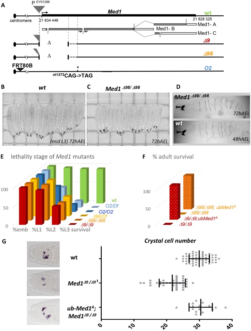
Drosophila Med1 is required for organism viability and hematopoietic crystal cell differentiation. (A) Med1 locus on chromosome 3 with the insertion site of the P element (EY01286; gray triangle) used for mutagenesis, the three Med1 alternatively spliced mRNAs (gray boxes) depicting the coding sequences, separated by intronic regions, and the molecular characterization of Med1 mutant alleles Δ9, Δ98, and O2. Deleted genomic sequences are represented by Δ. (B and C) Images of wild-type (wt) (B) versus Med1Δ98/Δ98 mutant (C) larvae at 72 h after egg laying (AEL). (D) A Med1Δ98 homozygous larva at 72 h AEL with a size equivalent to that of a 48-h AEL wt control and without external cuticular defects. (E) Complementation tests. Proportions of homozygous (Med1/Med1) or hemizygous (Med1/Df) Med1 mutant alleles dying at embryonic (emb) or larval stages L1, L2, and L3 are indicated. (F) Rescue tests. Proportion of ub-Med1A transgenic adults homozygous for Med1Δ9 or Med1Δ98 compared to the expected proportion of the F1 progeny for full rescue. (G) In situ hybridization of the crystal cell-specific PPO2 mRNA in wild-type, Med1Δ9/Δ9, or ub-Med1A; Med1Δ9Δ9 stage 14 embryos. Representative dorsal views of the embryo head region are shown on the left. The graph indicates the crystal cell number of each embryo of the given genotype.

