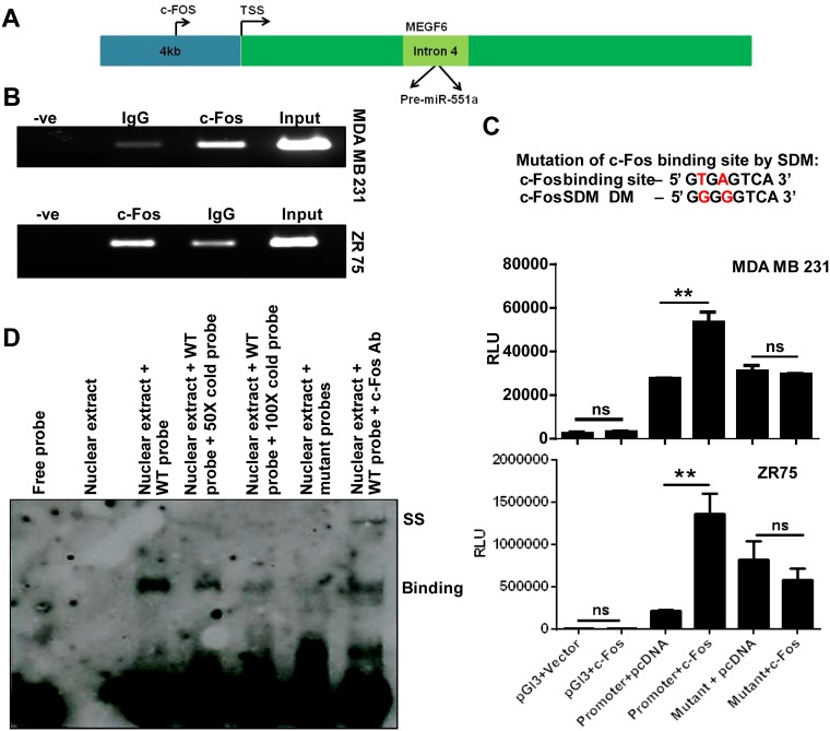
FIG 6.
c-Fos binds to the promoter of miR-551a. (A) Schematic showing the binding of c-Fos to the predicted binding site of the putative promoter region of miR-551a. (B) Representative ChIP images of agarose gel showing the PCR amplification for the putative promoter region of miR-551a upon immunoprecipitation with IgG or c-Fos antibody. (C) Bar graph representing the relative luciferase activity of cloned miR-551a promoter or mutant upon cotransfection with vector (pcDNA) or c-Fos (**, P < 0.05; ns, not significant, by one-way analysis of variance). (D) EMSA showing the binding of biotinylated wild-type (WT) probes (lane 3) spanning the c-Fos binding region in miR-551a promoter, competitive inhibition of band intensity with nonbiotinylated probes (lanes 4 and 5). The binding was abolished when biotinylated mutant probes were used (lane 6). The supershift was observed when the lysate was incubated with anti-c-Fos antibody (lane 7). −ve, negative (empty); RLU, relative light units; Ab, antibody; DM, double mutant.

