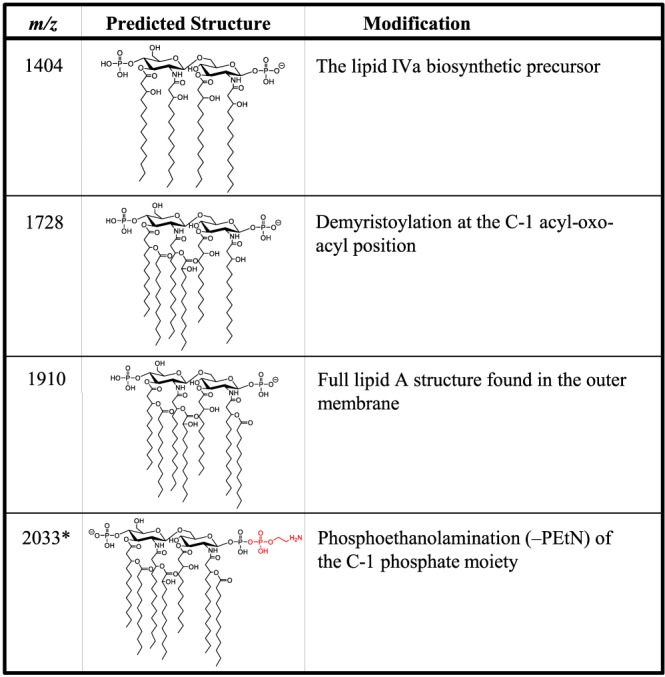
FIG 2.

Lipid A structures of signature ions with corresponding m/z values. The lipid A m/z values and the molecular structures found in the mass spectra of the A. baumannii clinical isolates with descriptions of the modifications responsible for the observed mass shifts are shown. Structures in red indicate modifications to the base structure at m/z 1,910. The charge position and location of phosphoethanolamine are arbitrary. *, ions associated with resistance to colistin.
