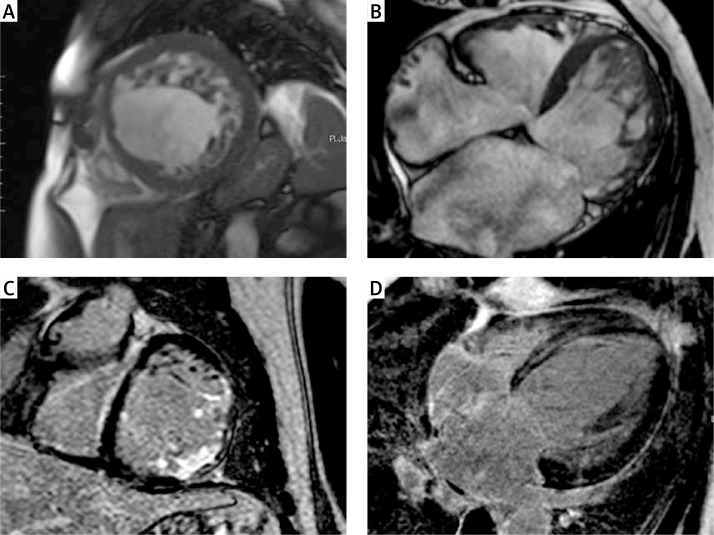
Figure 1.
Examples of cardiac magnetic resonance images of different patients with left ventricular non-compaction. Short axis (A) and four chamber (B) images with steady-state free precession (SSFP) sequences showing two layer structure with prominent trabeculations and deep intertrabecular recesses. Short axis (C) image of transmural and subendocardial late gadolinium enhancement in the basal anterolateral and inferolateral segments. Four-chamber (D) image of late gadolinium enhancement with mid-myocardial distribution in the interventricular septum

