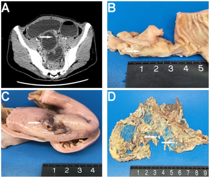
Figure 1.
(A) Enhanced computed tomography revealed a dilatation and wall thickening of the distal small intestine (arrowhead). (B) Macroscopically, a mass within the intestinal wall that was well-circumscribed and gray-white was identified (arrowhead). (C and D) Multiple gray-white nodules were found adhering to the serosa surface of the intestine and the omentum majus (arrowhead).

