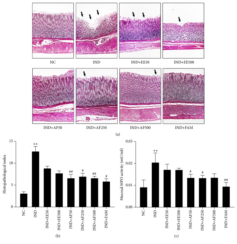
Figure 5.
Effect of pretreatment with EE and AF on histopathological observation of gastric mucosa. (a) Light micrograph of the gastric mucosal ulcer by IND (H&E, x100). Extensive disruption to the surface epithelium of gastric mucosa was indicated with a black arrow. (b) The histopathological index was calculated from the intensity of ulceration or erosion in the glandular epithelium, number of petechial, inflammatory cell infiltration, present of polymononuclear (PMN) cell, and expression of degeneration granular lining cell. (c) Mucosal MPO activity. The data are expressed ± SEM (n = 6) ∗∗P < 0.01 vs. NC group. #P < 0.05; ##P < 0.01 vs. IND group.

