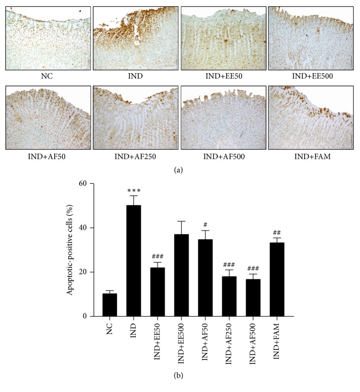
Figure 8.
Effect of pretreatment with EE and AF on apoptosis of gastric mucosa. Light micrograph of apoptotic cells (dark brown cells staining) in gastric ulcer induced by IND. The percentage of apoptotic-positive cells was analyzed using light microscopy with high power field (x400) from at least 5 fields. The data are expressed as mean ± SEM (n=6). ∗∗∗P < 0.001 vs. NC group. #P < 0.05; ##P < 0.01; ###P < 0.001 vs. IND group.

