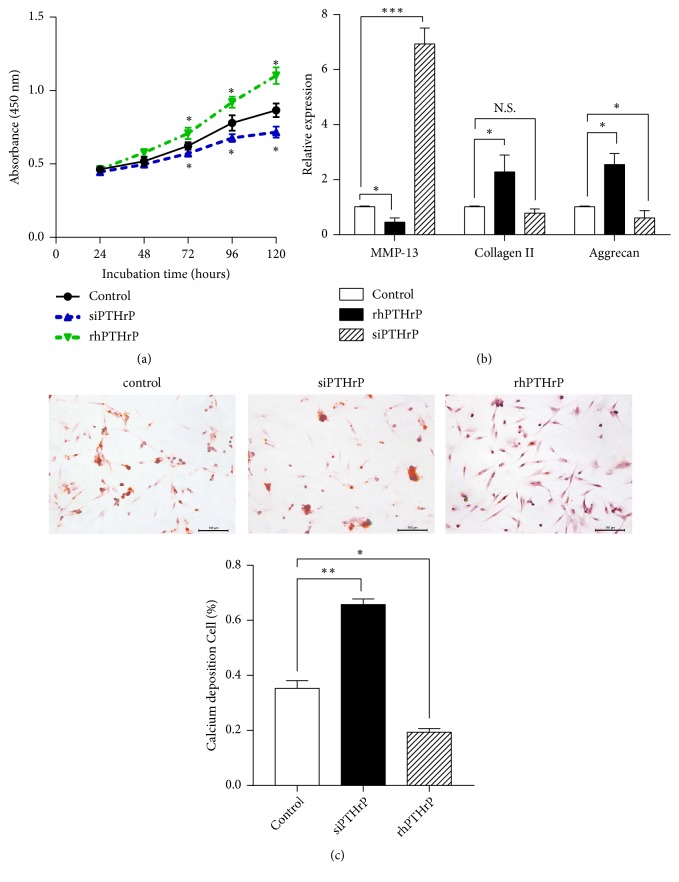
Figure 2.
Effect of PTHrP on chondrocyte proliferation and differentiation. OA chondrocytes were transfected with siPTHrP or a control plasmid or treated with rhPTHrP. (a) Cell viability was tested with a CCK8 kit after 24, 48, 72, 96, and 120 h. (b) qRT-PCR was performed to examine the MMP-13, collagen II, and aggrecan expression in the chondrocytes (∗p<0.05, ∗∗∗p<0.001 in comparison to the control, n = 6). (c) Calcium deposition was visualised by Alizarin Red S staining (scale bars, 100 μm). Calcium deposition (+) cells are presented as mean ± SD in the lower panels (∗p<0.05, ∗∗p<0.01, and n = 6).

