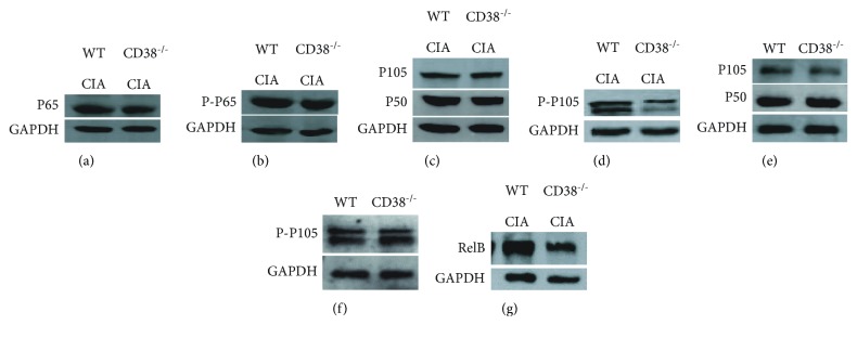
Figure 5.
Decreased phosphorylation of NF-κB in BMDCs generated over 7 days from CD38−/− CIA mice. The protein was extracted from DCs in the WT and CD38−/− mice with or without collagen stimuli. Expressions of NF-κB p65 (a), Phospho-NF-κB p65 (b), NF-κB1 p105 (c), Phospho-NF-κB1 p105 (d), and RelB (g) in WT CIA and CD38−/− CIA mice were detected by Western blot (n = 3 per group/experiment). Expressions of NF-κB1 p105 (e) and Phospho-NF-κB1 p105 (f) in the WT and CD38−/− healthy mice were also detected by Western blot as normal control (n = 3 per group/experiment).

