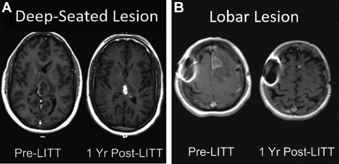
FIGURE 2.

Lesion locations. Pre- and postoperative imaging demonstrating successful radiographic response for tumors in A, left posterior thalamic and B, left frontal locations. Magnetic resonance-safe metal artifact is noted on the patient's right side in B.
