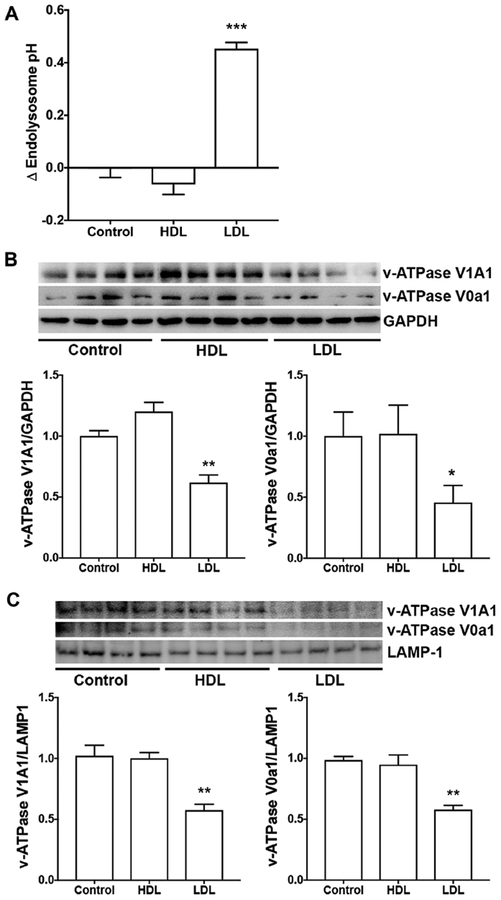
Figure 3. LDL, but not HDL, de-acidified endolysosomes.
(A) In SH-SY5Y cells expressing wild-type AβPP, LDL (50 μg/ml for 3 days), but not HDL (50 μg/ml for 3 days), significantly elevated endolysosome pH (***p<0.001, n=16–20 cells from 4 different plates). (B) LDL, but not HDL, significantly decreased protein levels of V1A1 and V0a1 subunits of v-ATPase in total cell lysates of SH-SY5Y cells (*p<0.05, **p<0.01, n=4). (C) LDL, but not HDL, significantly decreased protein levels of v-ATPase V1A1 and V0a1 subunits in enriched lysosome fractions of SH-SY5Y cells (**p<0.01, n=4).

