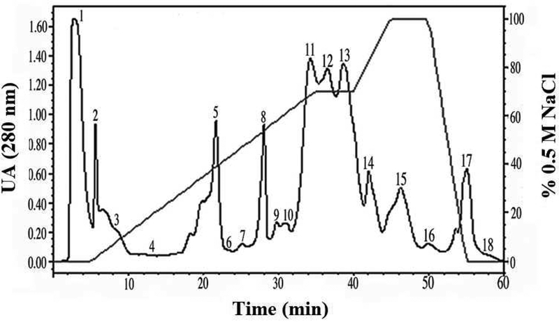
Fig. 1. Cation-exchange chromatography from A. c. pictigaster venom.
Five hundred microliters of A. c. pictigaster venom (60 mg/mL) was injected to a cationic exchange HPLC column Waters™ SP 5PW (75 × 7.5 mm). The fractions were separated with a 0.02 M sodium phosphate buffer, pH 6.2, containing 0.5 M NaCl. The separation required 60 minutes with a flow rate of 1.0 mL/min. The absorbance was measured at 280 nm by a Waters™ 2487 Dual absorbance detector. The proteolytic activity was tested (hemorrhagic, fibrinolytic, and gelatinase) for each fraction.

