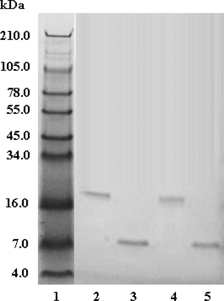
Fig. 3. SDS-PAGE analysis of A. c. pictigaster venom fractions from C18 HPLC column.
Venom fractions (20 μg) were run on 10–20% Tricine SDS-PAGE under non-reducing and reducing conditions at 125 V for 90 min. The gel was stained with Simply Blue Safe Stain for 1 h and distained overnight with Milli-Q water. . Lane 1: SeeBlue Plus2 Markers (Invitrogen™); Lane 2: pictistatin 1 (non-reduced); lane 3: pictistatin 1 (reduced); lane 4: pictistatin 2 non-reduced; lane 5: pictistatin 2 reduced. (reduced)

