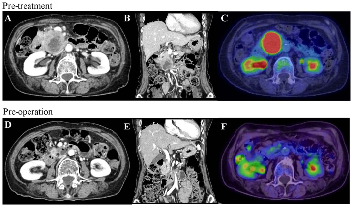
Figure 1.
CT and PET before and after gemcitabine plus nab-paclitaxel therapy. (A) CT at the initial visit showed a hypovascular tumor measuring 50 mm in the head of the pancreas. The tumor was in contact with the SMA. (B) Tumor invasion extended to the most proximal draining jejunal branch into SMV and the tumor spread over one third of the duodenum. (C) PET at the initial visit showed high fluorodeoxyglucose uptake into the primary pancreatic tumor. (D) Tumor size decreased to 18 mm post-operation, contact with the SMV decreased to 90 degrees and the tumor separated from the SMA. (E) Tumor invasion in proximal draining jejunal branch into the SMV disappeared. (F) Fluorodeoxyglucose uptake into the primary pancreas tumor was eliminated. CT, Computed tomography; PET, positron emission tomography; SMA, superior mesenteric artery; SMV, superior mesenteric vein.

