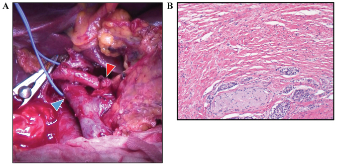
Figure 3.
Intraoperative and pathological images. (A) Intraoperative image. Gastroduodenal artery stump is indicated by the red arrowhead and the portal vein is indicated by the blue arrowhead. (B) Microscopic findings, following chemotherapy, of the surgical specimen showing a change of <10% in the fibrous tissue with grade I on Evans' grade criteria. Staining was performed with hematoxylin and eosin (magnification, ×40).

