Fig. 2.
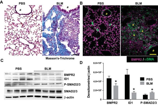
Bone morphogenic protein receptor 2 (BMPR2) expression is depleted in experimental lung fibrosis. Masson's trichrome (A) or immunofluorescently (IF) stained lung sections (B) from BMPR2 (violet/purple signals) and α-smooth muscle actin (αSMA/green signals) from phosphate-buffered saline (PBS)- or bleomycin (BLM)-exposed mice, showing increased fibrotic deposition in BLM-exposed mice. A, Airway structures; asterisks identify fibrotic areas in the lung. Scale bar = 100 μm. C: immunoblots for BMPR2, ID1, P-SMAD 2/3, and β-actin from PBS- and BLM-exposed mice. D: densitometries for BMPR2, ID1, and P-SMAD2/3. *P ≤ 0.05.

