Fig. 5.
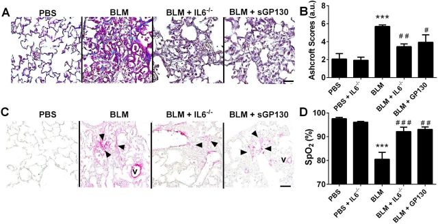
Blocking IL6 attenuates lung fibrosis. Masson's trichrome stain (A), Ashcroft scores (B), IHC (C) for smooth muscle actin (αSMA/magenta signals) and counterstained with methyl green. V, vessel, arrowheads denote myofibroblasts; from PBS, BLM, BLM+IL6−/− and BLM+sGP130 treatment groups. D: arterial oxygenation (SpO2) determined by pulse oximetry from PBS, BLM, BLM+IL6−/−, and BLM+sGP130 groups. ***P ≤ 0.01, ANOVA comparisons between PBS and experimental treatment groups. ###P < 0.001, ##0.001 < P < 0.01, #0.05 < P, ANOVA comparisons between BLM and BLM+IL6−/− or BLM+sGP130 treatment groups.

