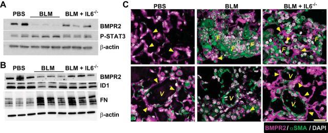
Fig. 6.
BMPR2 signals are maintained in IL6-deficient mice. Immunoblot for BMPR2, P-STAT3, and β-actin (A); immunoblot for BMPR2, inhibitor of differentiation (ID) 1, fibronectin (FN), and β-actin (B) from PBS, BLM, and BLM+IL6−/− treatment groups. C: immunofluorescence for BMPR2 (magenta signals) or α-smooth muscle actin (αSMA/green signals) and counterstained with DAPI (white/gray signals) from PBS, BLM, and BLM+IL6−/− treatment groups. Arrowheads denote signals for BMPR2. V, vessel; F, fibroproliferative lesions. Scale bar = 50 μm.

