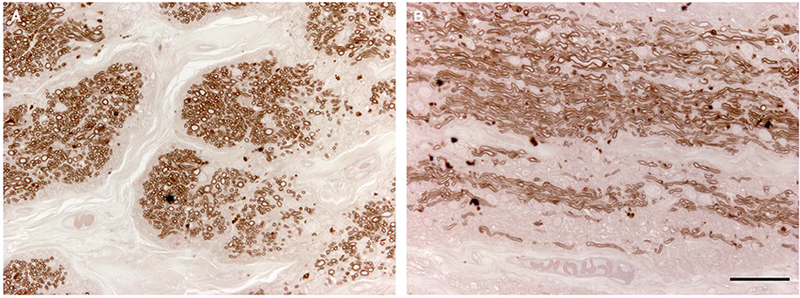
Figure 3. Histologic features of the optic nerve of an eye with a MYOC.

Tyr437His mutation. The myelin sheaths of optic nerve axons were stained with paraphenylenediamine (PPD). Extensive loss of axons caused by severe glaucoma is seen in cross-section (A) and sagittal section (B) of patient AR’s optic nerve. Scale bar = 100μm.
