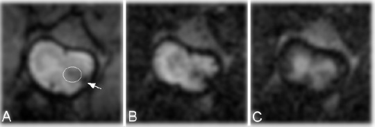
Figure 4.

Magnetic resonance images from the representative injured spinal cord after ebselen treatment at 12 weeks.
(A) T2-weighted image. Diffusion-weighted image with diffusion gradient applied along phase direction (B) and (C) slice direction. Scar/hemorrhage pathological change present in the white matter (indicted by arrow) and at the border between the white and gray matter of lateral funiculus of spinal cord (marked with the circle).
