
Figure 6.
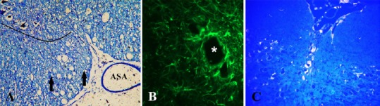
Microphotographs obtained 12 weeks after spinal cord injury in rats treated with ebselen.
(A) Spinal cord, toluidine blue staining; numerous cystic cavitations (vacuolar structures). Lined area of anterior horn of spinal cord; ASA: anterior spinal artery; arrows: axon spheroids (swollen axons). (B) Formation of glial fibrillary acidic portein primary-labeled cavity in the injured spinal cord ventral white column. Asterisk: microcyst with astrogliosis. (C) FluoroGold retrograde labeling of regenerating neurons. Neuronal cytoplasm is labeled by fluorogold in transverse sections of the raphe nuclei in the brain stem.