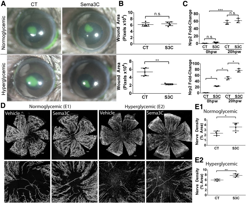
Figure 5.
Exogenous SEMA3C (S3C) increases DM wound healing and corneal nerve regeneration in healing corneas. NL and DM corneas pretreated with BSA as the control (CT, right eyes) or recombinant S3C (50 ng/μL, left eyes) 4 h before epithelium debridement. At 0 h, the corneas were wounded by epithelium debridement (1.5-mm diameter) and allowed to heal for 20 h (A–C) or 3 days (D and E). A: At 20 hours postwounding (hpw) in NL mice and 24 hpw in DM mice, injured corneas were stained with FITC to photograph the remaining wound area. B: The wound sizes were calculated using Adobe Photoshop software and presented as the average of total pixels (mean ± SD, n = 5). **P < 0.01 (Student t test). C: CECs collected during wounding (20 hpw) and from original wound beds (0 hpw) were subjected to RNA qRT-PCR analysis of Nrp2 expression. The results are presented as fold increase (mean ± SD) over the NL nonwounded CECs, which were set as a value of 1 (n = 3). *P < 0.05, ***P < 0.001 (two-way ANOVA). D: Representative corneas harvested at 3 days postwounding (dpw) for NL and 4 dpw for DM, with an additional subconjunctival injection of BSA or recombinant S3C at 1 dpw. Staining for β-tubulin III was performed, and images of the entire cornea were captured (top panels), with high-magnification images shown in bottom panels. Scale bars: 176 μm (E1), 202 μm (E2). E1 and E2: Nerve densities at the central areas (bottom panels) were calculated using ImageJ software from the areas covered with β-tubulin III staining and presented as percent area (mean ± SD, n = 4). Two independent experiments were performed. *P < 0.05, **P < 0.01 (Student t test). n.s., not significant.

