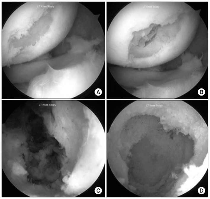
Fig. 3.
Arthroscopic view of the osteochondral lesion. (A) Full thickness cartilage defect over the weight bearing portion of lateral femoral condyle as seen from the anteromedial portal. (B) Breakdown of the subchondral bone upon probing the base of the cartilage defect. (C) The interior of the bone cyst as seen by the arthroscope inserted from the anterolateral portal revealed hemorrhagic tissue within the cavity. (D) The interior of the cyst as seen after complete curettage of the cavity.

