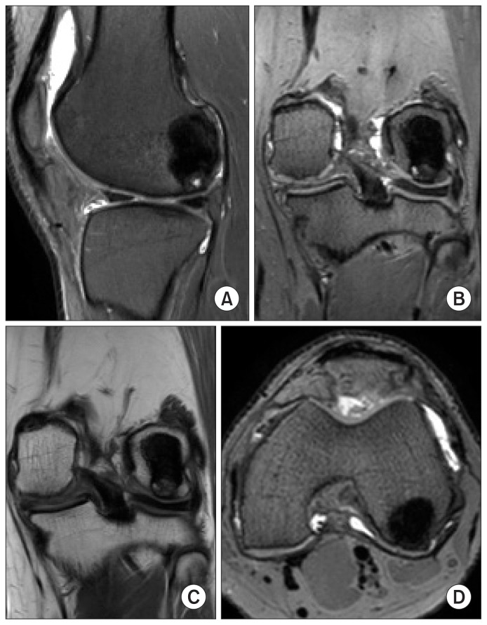
Fig. 5.
Postoperative magnetic resonance imaging (MRI) at the final follow- up: the bone defect has completely consolidated and the implanted osteochondral plug is well incorporated with no surrounding edema as seen on the proton density sagittal (A), proton density coronal (B), T1- weighted coronal (C) and proton density axial (D) images.

