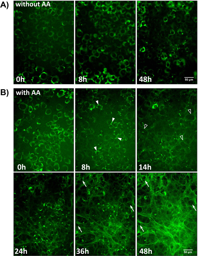
Figure 5: Time Lapse Imaging of collagen assembly in MLO-colGFP cells.

A) Still frame images from a timelapse movie showing MLO-colGFP cells imaged without ascorbate (without AA). Note that the GFP-collagen remains intracellular and there is no deposition of extracellular fibrils (see supplementary movie 1). B) Still frame images from a timelapse movie showing MLO-colGFP cells imaged with ascorbate (with AA) (see supplementary movie 2). Note that after ascorbate addition at 0h, the GFPtpz-collagen migrates into vesicle-like structures by 8h (arrowheads) and that faint GFPtpz-positive fibrils are seen by 14h (open arrowheads). An extensive collagen fibril network is assembled by 48h. The cells appeared to physically reshape the collagen matrix by pushing collagen fibrils outwards to generate small holes in the fibril network (compare arrows at 36h and 48h). Bar = 50μm. [Still images in A) & B) are representative of >3 experiments, including >40 movies with AA and >15 movies without AA]
