
Figure 6: ECM and Cell Dynamics During Collagen Assembly.
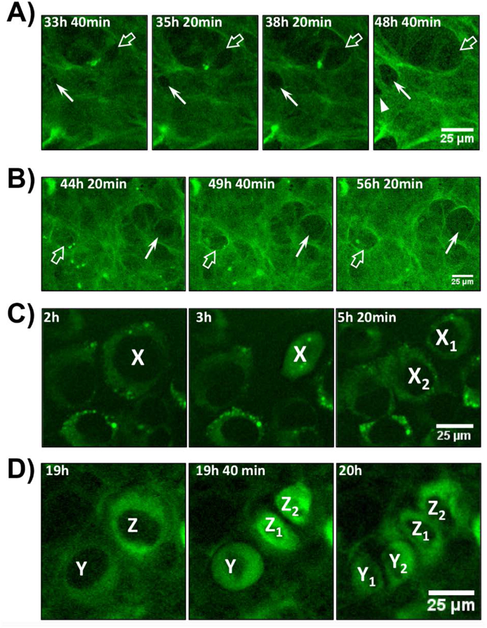
A) Enlarged view of still frame images from movie 2 showing MLO-colGFP cells imaged with ascorbate. Note the appearance of two small holes in the collagen fibril network at 33h 40 min (arrow and open arrow), which enlarge over successive image frames. An additional hole (arrowhead) appears at 48h 40 min. B) Enlarged view of still frame images from movie 2 showing another example of the formation of holes in the GFP-collagen fibril network that enlarge over time (arrow and open arrow). C) Enlarged view of still frames from movie 1 showing MLO-colGFP cells imaged without ascorbate. A cell, X, starts dividing at 3h and shares its GFPtpz-collagen content equally between daughter cells, X1 and X2. D) Enlarged view of still frames from movie 2 showing MLO-colGFP cells imaged with ascorbate. A cell, Z, starts dividing after 19h and shares its collagen content equally between daughter cells, Z1 and Z2. A second cell, Y divides by 20h and also shares its collagen content equally between daughter cells Y1 and Y2. Bar in A-D = 25μm. [Images are representative of >3 experiments (>20 movies) in A) & B) and >3 experiments (>15 movies) in C) & D)]