
Figure 9. TS aorta SMAD staining.
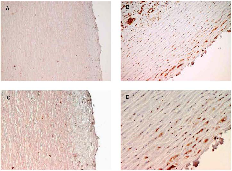
Immunohistochemical analysis the same aortas shown in figure 6 to assess levels of TGF-β signaling. The two left panels (A, C) are from control aortas. The two right panels are sections from the TS aorta. All are stained with p-SMAD antibody. SMADs are downstream products of TGF-β signaling. Note the increase in staining (reddish-brown) in the TS aorta (Panels B and D) compared to a normal control (Panels A and C), indicating an increase in p-SMAD production as a result of increased TGF-β signaling. Excessive TGF-β signaling is seen in aortas from individuals with Marfan syndrome and is thought to contribute to aortic wall instability. This is the first demonstration of excessive TGF-β signaling in TS.