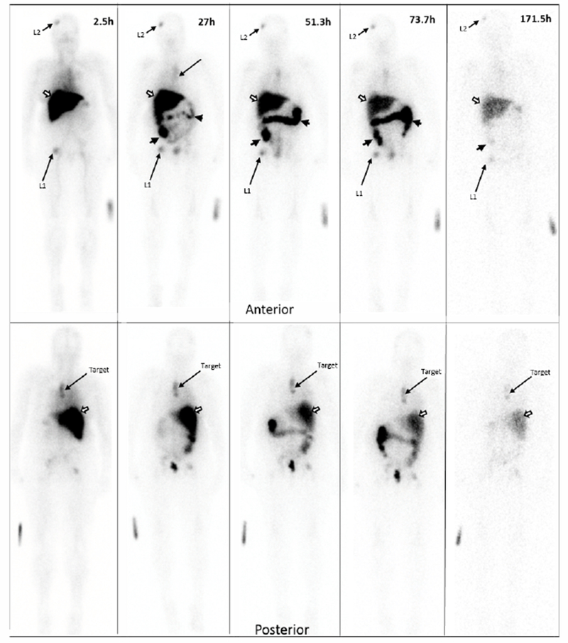
Figure 2A.

Planar imaging of a HER2
True Positive patient over time. Serial Anterior (top row) and Posterior (bottom row) Planar imaging of a breast cancer patient following the i.v. administration of 186.8 MBq (69.7μg) of 111In-CHX-A”-DTPA trastuzumab (23.7-minute wholebody acquisition, 1.3mm/s, 1.85m). The HER2(+) left paraspinal mass (“target” on the bottom row) shows focal 111In-CHX-A”-trastuzumab uptake. Other prominent foci in the right frontal bone (L1) and right hip (L2) are consistent with patient’s known metastatic bone disease. The remaining distribution is physiologic, seen throughout all patients: accumulation of tracer in the liver (white arrow) and large bowel (black arrow heads) and no significant brain or cardiac uptake. Times indicate time post-injection. Images are scaled to represent biological changes only. Radioactive decay is NOT depicted. Degradation of imaging quality at the 171.5h time point is due to low counts.
