
Figure 3B.
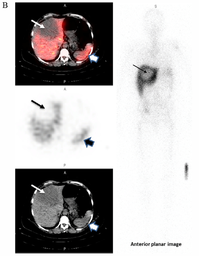
True negative.
This 56-year-old female with metastatic breast cancer presented with a large hypodense liver lesion (narrow arrow on left) seen best on the low dose CT scan (bottom image). Biopsy showed no evidence of HER2 overexpression (HER2 1+; FISH 1.6). SPECT images performed at 173h post-infusion of 189.8MBq[136.5μg]. 111In-CHX-A”-DTPA trastuzumab shows no significance uptake compared to liver background (top SPECT and middle fused SPECT/CT image). Lesion to Background ratio is 0.85. The wider arrow on the right indicates the spleen. Some mis-registration is present likely due in part to respiratory motion