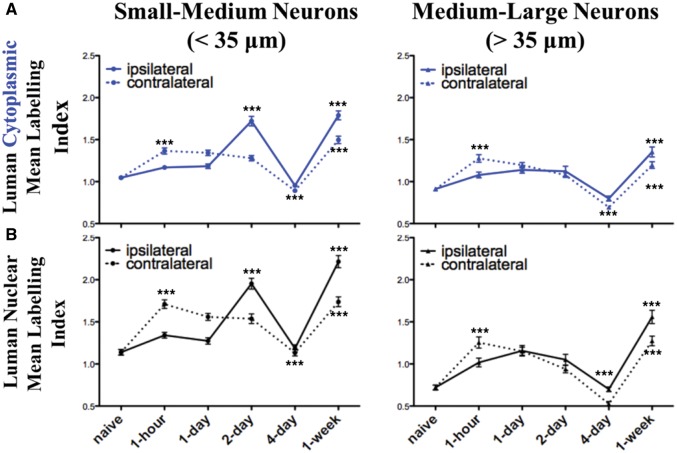
FIGURE 4.
Peripheral unilateral axotomy results in bilateral biphasic alterations in Luman protein immunoreactivity in both injured and contralateral uninjured dorsal root ganglion (DRG) neurons. Summary line graphs of alterations in the mean + SEM. cytoplasmic (A, blue) and nuclear (B, black) Luman immunofluorescence intensity levels as normalized to the mean naïve control value on the same slide. Data were subdivided into that observed in small- and medium-sized (<35 μm, column 1) and medium- and large-sized (>35 μm, column 2) DRG neurons ipsilateral (injured—solid line) and contralateral (uninjured—dashed line) to injury at timepoints as indicated (n = 181–206 neurons analyzed per timepoint per animal analyzed), with 3 rats/experimental condition assessed for each data point. ***p < 0.001 ANOVA with Dunn’s post-test analysis for change relative to previous timepoint. Note: relative changes in nuclear localization parallel those observed for the cytoplasmic staining.

