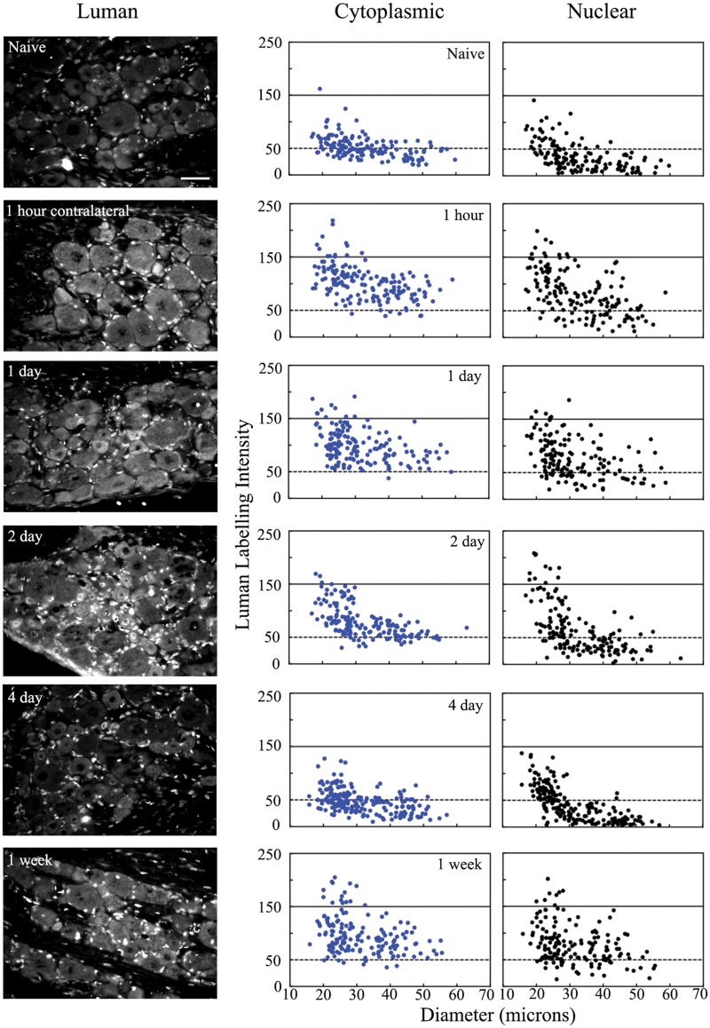
FIGURE 5.
Unilateral peripheral nerve injury alters Luman protein levels in uninjured dorsal root ganglion (DRG) neurons contralateral to injury. Left Column: Representative photomicrographs of L5 DRG sections (6 μm) processed for immunofluorescence to detect Luman protein in DRG contralateral to 1-hour, 1-day, 2-day, 4-day and 1-week injury. Sections shown were all mounted on the same slide to ensure processing under identical conditions and are those for which the quantification is shown. Scale bar = 50 μm. Naïve animals served as controls. Right Column: Representative scatterplots depicting relative changes in Luman immunofluorescence signal over neuronal cytoplasmic and nuclear regions as related to neuron size. Experimental states as indicated. Dashed lines divide the plots into low versus moderate to heavily labeled populations (n = 181–204 neurons analyzed per animal per condition). A total of 3 separate animals were analyzed in this manner per condition.

