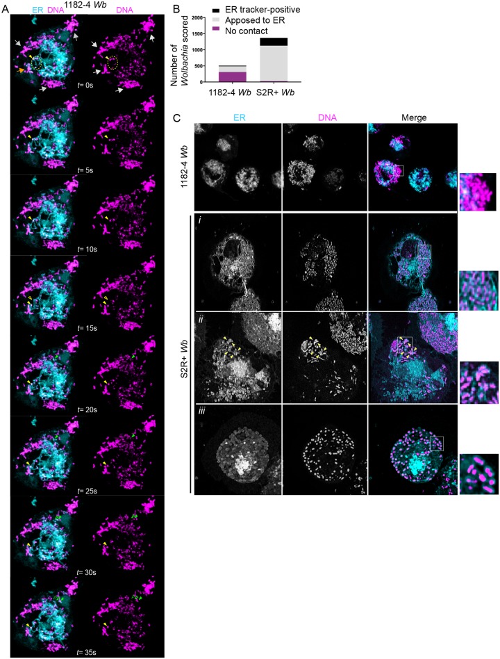
Fig 3. Wolbachia physically interact with the endoplasmic reticulum.
(A) Time-lapse acquisitions at a surface focal plane in a 1182–4 Wb cell stained with the DNA dye SYTO 11 -magenta- to highlight Wb, and an ER tracker -cyan-. A t = 0 second, grey arrows point to peripheral Wb clusters that are not in close contact with the ER. The orange arrow points towards some Wb remaining in close contact with the ER during the time lapse duration. The dotted yellow circle highlights some Wb located within ER tubules. A single Wb within an ER tubule is tracked by the plain yellow arrowhead, and its previous position is indicated by an empty yellow arrowhead (i.e. at t = 15s). Similarly, the movement of a single Wb surrounded by an ER-derived membrane is tracked by green arrowheads (t = 15s to t = 35s). See the corresponding supplemental movie 3. (B) Scoring of Wb- ER interactions, observed with SYTO 11 and the ER tracker in 1182-Wb and S2R+ Wb cells, in random focal planes of n = 18 and n = 12 cells respectively. Bacteria co-localized with ER tubules, or surrounded by an ER tracker-positive membrane were counted as "ER-tracker positive". (C) The different interactions between Wb and the ER are highlighted on these confocal images, with clusters of Wb not in contact in 1182–4 Wb -see inset-. The following rows are different examples in S2R+ cells showing i) Wb in close contact with the ER, ii) a Wb cluster composed of individual Wb surrounded with an ER tracker-positive membrane -yellow arrowheads-; iii) and in rare instances all individual Wb of the cell being surrounded with an ER tracker-positive membrane.

