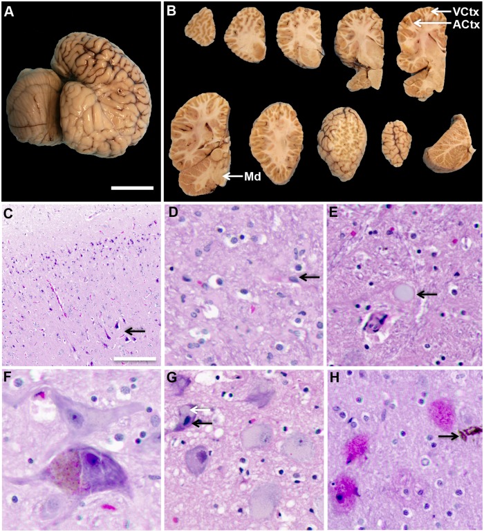
Fig 2. Gross and microscopic evaluation of postmortem brains from stranded dolphins.
(A) External examination was performed on the cerebral cortex and cerebellum of formalin-fixed hemispheres from stranded dolphins (n = 7). (B) Following external examinations, brain hemispheres were cut into a series of coronal slices to investigate internal gray and white matter structures. Tissue blocks were sampled from anatomical regions in the dolphin cerebral cortex and brainstem involved with acoustico-motor navigation: auditory cortex (ACtx), visual cortex (VCtx), and the medulla oblongata (Md). (C) Digital pathology scans were obtained from routine histological stain. H&E stain shows hypoxic and eosinophilic changes in neurons of both upper and lower cortical layers. (D) Gliosis was also observed in the cerebral cortex. (E) Advanced age-related changes were observed including, corpora amylacea and (F) lipofuscin granules. (G) Karyorrhexis nuclear changes (black arrow) and chromatolysis (white arrow) were observed. (H) Representative scans of eosinophilic plaques and a rare hemosiderin deposits were observed in the ACtx of stranded dolphins. Representative scale bar: 5 cm (A, B), 1000 μm (C), 200 μm (D, F, G), 50 μm (E, H).

