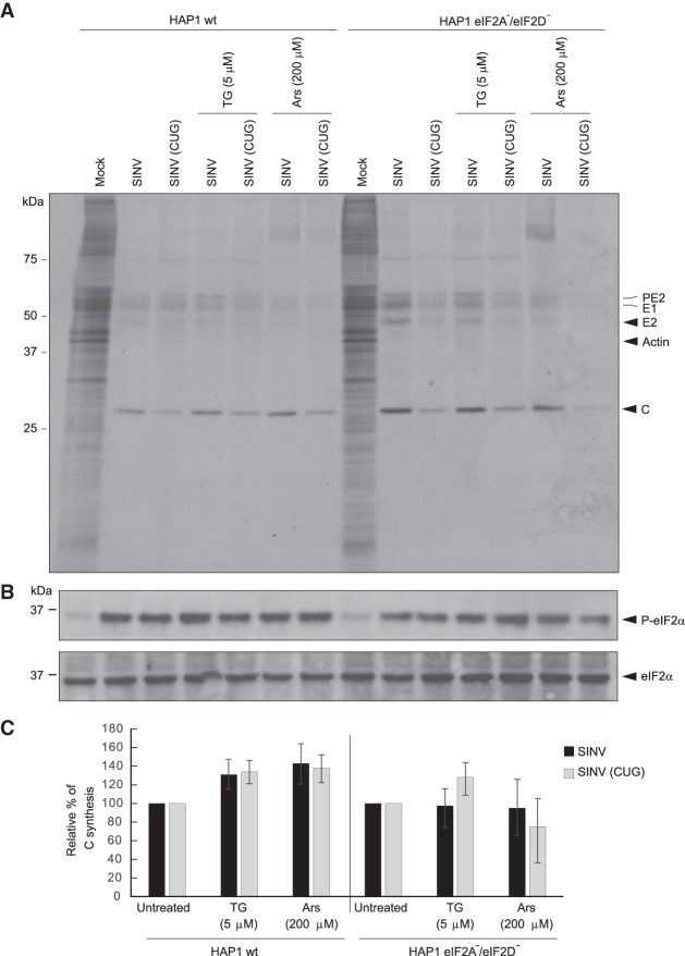
FIGURE 9.
Infection of HAP1 wild-type (wt) and the double KO cell line HAP1 eIF2A−/eIF2D− by wt SINV or SINV CUG. (A) HAP1 wt and HAP1 double KO cells were mock infected or infected with 10 plaque-forming units/cell wt SINV or SINV (CUG) for 1 h. Then, the infective medium was replaced by fresh growth medium. At 7 h post-transfection, cells were incubated in radioactive labeling medium with [35S] Met/Cys and treated or not with thapsigargin (TG; 5 µM) or sodium arsenite (Ars; 200 µM) for 1 h. Then, cells were collected in loading buffer and analyzed by SDS-PAGE, fluorography and autoradiography. (B) In parallel, the state of phosphorylation of eIF2 was analyzed by western blotting with anti-P-eIF2α and anti-eIF2α antibodies. (C) Densitometric analysis of C synthesis is shown in the graphs as relative to their corresponding untreated samples. The results are displayed as mean ± SD of three representative experiments. The black bars correspond to translation initiation at AUG and gray at CUG.

