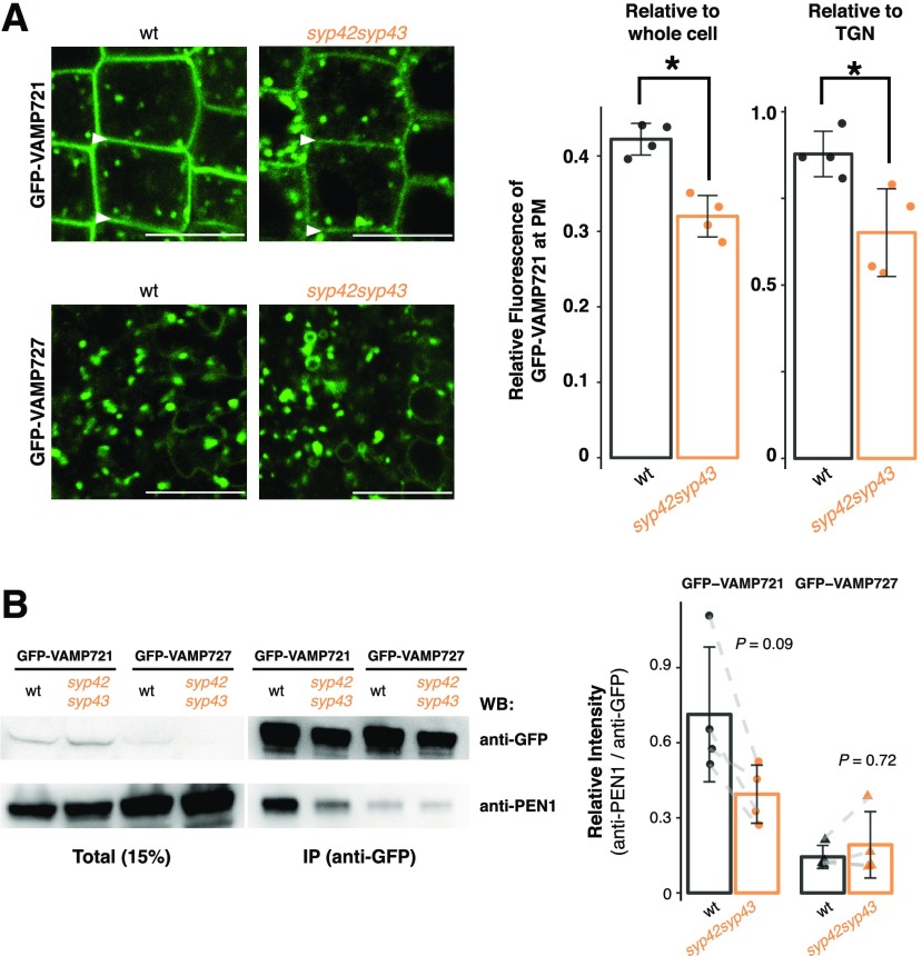
Figure 2.
The R-SNARE VAMP721 operates in the SYP4-dependent secretory pathway. A, Representative confocal images of Arabidopsis roots expressing GFP-VAMP721 and GFP-VAMP727 in the wild-type (wt; left) and syp42syp43 (right) mutant plants. White arrowheads indicate the PM. The graph shows the relative fluorescent intensity of GFP-VAMP721 at the PM compared with whole cell (left; n = 8) or to TGNs (right; n = 8) in the wild-type and syp42syp43 mutant plants. B, Protein extracts of Arabidopsis shoots expressing GFP-VAMP721 and GFP-VAMP727 in the wild-type and syp42syp43 mutant plants were immunoprecipitated with anti-GFP antibody and detected with anti-PEN1 and anti-GFP antibodies by western blotting. The graph shows signal intensities of coimmunoprecipitated PEN1 normalized by the signals from GFP-VAMP721 or GFP-VAMP721 (n = 14). Asterisks in (A) indicate statistical significance based on Student’s t test (α = 0.05). P values in (B) calculated using Student’s two-sample paired t test with Bonferroni correction for multiple comparisons (α = 0.05). Bar plots are shown with mean ±sd and raw data points. Scale bars = 10 µm.

