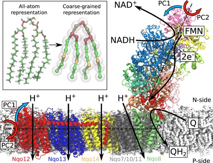
Fig. 1. Structure and function of the respiratory complex I (PDB ID: 4HEA).

Electrons are transferred from the NADH/FMN (flavin mononucleotide) site to quinone (Q) via a tunneling wire comprising eight iron-sulfur centers. The Q reduction takes place at the interface between the hydrophilic and membrane domains and couples to the pumping of four protons across the membrane. The global bending (PC1, blue arrows) and twisting motions (PC2, red arrows) are shown along two principal axes (see text). Inset: Comparison of atomistic and coarse-grained molecular simulation models of a cardiolipin molecule.
