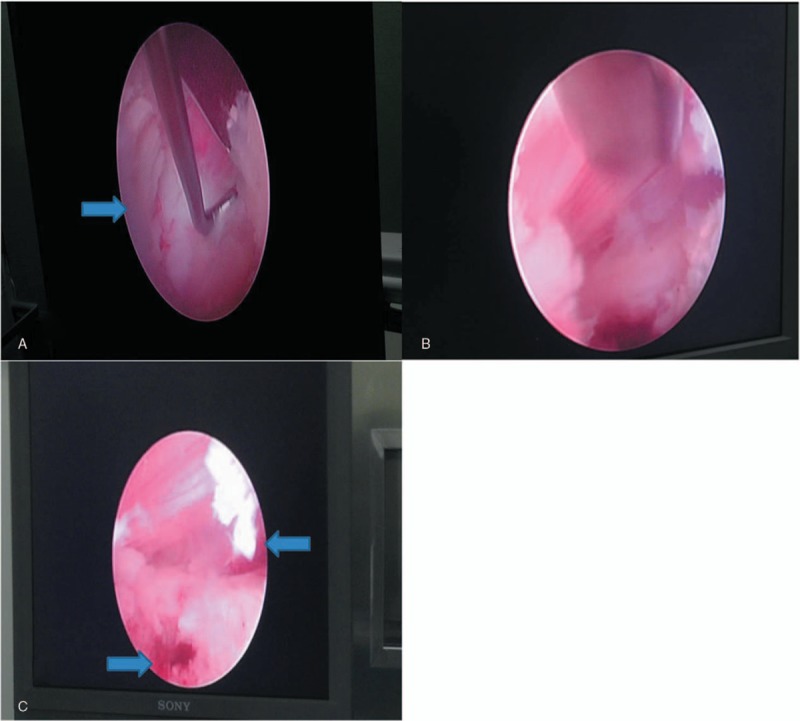
Figure 2.

A: The tumor compressed the dural sac and nerve root (Arrow). B: When part of the tumor tissue was exposed, we grasped and removed it with endoscopic forceps. C: After complete decompression, the dural sac and the L4 nerve root were lax in the endoscopic vision (Arrow).
