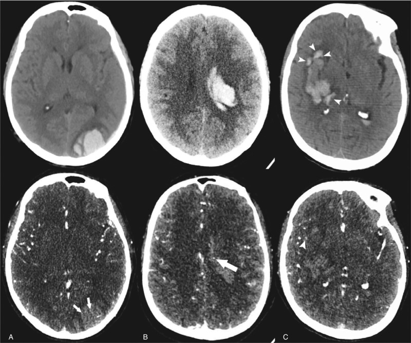
Figure 1.

Illustration of spot sign and island sign. (A) Unenhanced CT shows an intracerebral hemorrhage of left occipital lob, and the CTA demonstrates spot signs presenting as enhancement foci within main hematoma (arrow) (75-year old female). (B) A 54-year old individual presenting a mild hemorrhage and CTA imaging shows a spot sign within the main hematoma (arrow). (C) A right basal ganglia hemorrhage was found in a 63-year old male patient. The hematoma consists of 4 separate adjacent small hematomas which were identified as island sign (arrowheads) in unenhanced CT. Note that a spot-like foci of enhancement which is located around a small hematoma but not within the main hematoma is NOT considered as spot sign (arrowheads). CTA = computed tomography angiography.
