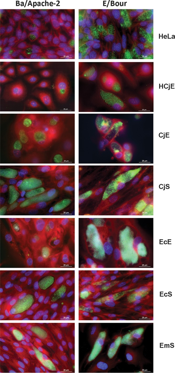
FIG 2.

Representative images of inclusion formation by C. trachomatis reference strains Ba/Apache-2 and E/Bour. CjE, CjS, EcE, EcS, and EmS cells and immortalized HeLa 229 and HCjE cells were infected simultaneously with Ba/Apache-2 or E/Bour at an MOI of 1. The same medium was used for all infections. The cells were grown on 24-well glass-bottom plates, fixed and stained at 48 hpi, and imaged using a Nikon Eclipse Ti-E inverted microscope with an LED illumination system and a DS-Qi2 camera at ×90 magnification. Representative images are shown from more than three independent experiments.
