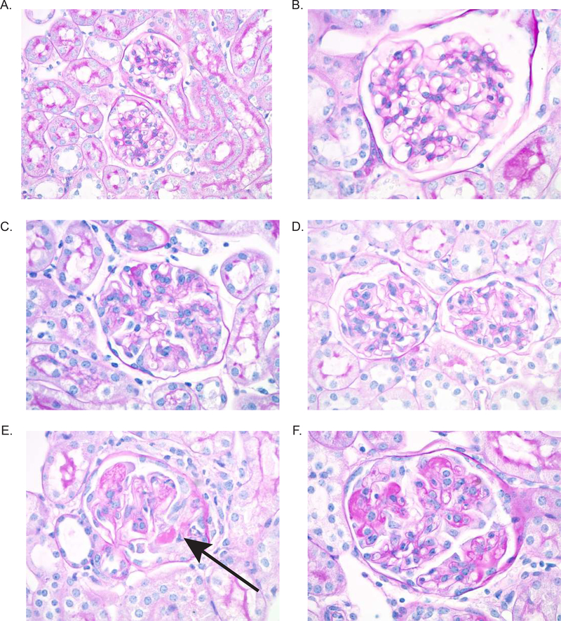
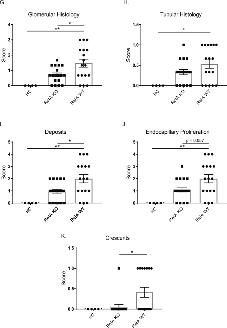
Figure 2. Renal histology.
Kidney sections were stained with PAS, and images were taken at 600x. Representative images were selected from HC (A and B), KO (C and D), and WT (E and F) mice. WT mice showed significant crescent formation (E, arrow), as well as endocapillary proliferation and immune deposits (F). Myeloid RelA KO mice, however, displayed much more normal histology, despite minimal endocapillary proliferation (C and D). There was significant improvement in the glomerular histology in KO mice (G), but neither group had a significant degree of tubular disease (H). Specifically, KO mice showed improvements in deposits, endocapillary proliferation, and crescents (I-K). Sections from 3 separate cohorts were scored by a renal pathologist blinded to the genotype (HC, n=4; RelA KO, n = 18; RelA WT, n = 17). Normally distributed data (glomerular histology, deposits, and endocapillary proliferation) were analyzed using an ordinary one-way ANOVA, and multiple comparisons were determined using Tukey’s test. Nonparametric data (Tubular histology and crescents) were analyzed using a Kruskal-Wallis test, and multiple comparisons were assessed with Dunn’s test.


