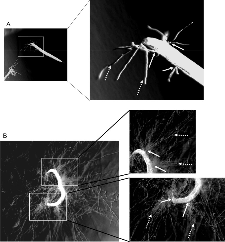
FIG 7.
Images of C. elegans AU37 with hyphae of F. oxysporum ISS-F4 piercing through worm body. Microscopic images taken at 22 h (A) and 46 h (B) after coincubation of C. elegans with ISS-F4 conidia. Solid arrows point out the hyphae piercing through the worm body. Dotted arrows show growing extended hyphae that initially penetrated from the intestinal tube. The images were taken with the Olympus SZX16 microscope (×5 magnification).

