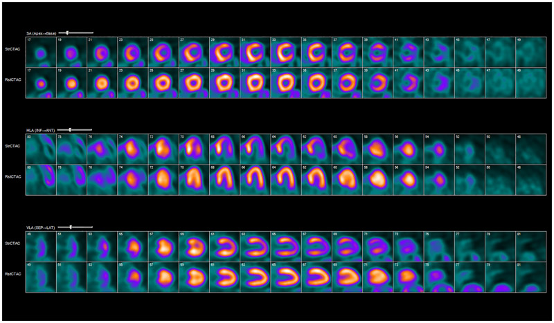
Figure 1:
Rubidium-82 PET myocardial perfusion imaging. Example of abnormal myocardial rest/stress perfusion imaging with rubidium-82 PET. Coregistered rest and stress images show a large reversible defect in the apex, apical anterior, and lateral walls consistent with ischemia. The patient had drop in ejection fraction and evidence of transient ischemic dilatation of the left ventricle with stress. StrCTAC = stress computed tomography attenuation correction; RstCTAC = rest computed tomography attenuation correction; HLA = horizontal long axis; VLA = vertical long axis; SA = short axis; INF = inferior; ANT = anterior; SEP = septal; LAT = lateral.

