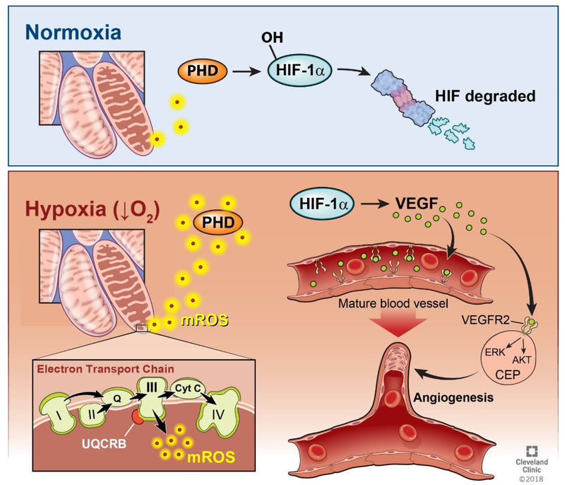
Fig. 1. Summary of the Angiogenesis Pathway.
Ischemia, exercise, and inflammation create a hypoxic tissue environment, resulting in decreased oxygen availability in cells. Under these hypoxic conditions, the electron transport chain produces mROS at mitochondrial Complex III. These mROS exit the mitochondria and deactivate prolyl hydroxylase (PHD). Under normoxic conditions, the levels of mROS being produced are insufficient to deactivate PHD, and PHD therefore hydroxylates the HIF-1α protein, marking it for eventual degradation by proteasomes. When high levels of mROS are produced under hypoxia, HIF-1α is stabilized, allowing the production of the VEGF protein. This VEGF protein attaches to the VEGFR2 receptor on both mature endothelial cells lining blood vessels and circulating endothelial precursors (CEP). This activates the ERK and Akt pathways, causing the maturation and mobilization of endothelial cells, allowing angiogenesis to occur.

