Figure 4.
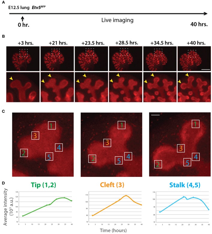
Expression pattern of ETV5-RFP in distal lung epithelium during early development (A) Experimental design: E12.5 Etv5RFP lung explants were cultured for 40 h and live imaged. (B) Still images showing representative ETV5-RFP expression both globally and in the distal tips (boxes). Arrows indicate increased expression in the tips over time. Scale bar: (Top row) 500 μm; (Bottom row) 125 μm. (C) Example images of a branching tip at three successive time points (a–c), highlighting three regions of dynamic ETV5-RFP expression: the tip (1 and 2), the cleft (3), and the stalk (4 and 5). See text for details. Scale bar: 37 μm. (D) Representative plot of ETV5-RFP expression in three independent regions [(a) tip, (b) stalk and (c) cleft] of a single bud over a period of 40 h (n = 1; a.u. = arbitrary units).

