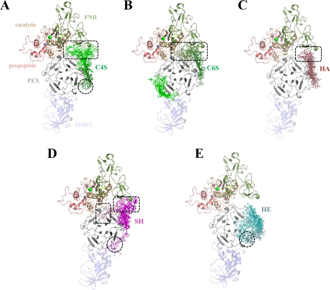
Figure 5.
Molecular modeling of GAG in complex with proMMP2. Docking results using Autodock3 and DBSCAN clustering are shown. proMMP2 (PDB ID 1GXD (3.1 Å)) is shown in cartoon and colored by domains: propeptide (light pink), catalytic domain (pale), FNII (green) and PEX domain (grey). Different clusters of GAG are shown in sticks with gradient colors: (A) C4S (green), (B) C6S (green), (C) HA (brown), (D) SH (pink) and (E) HE (teal). Calcium and zinc ions are shown in green and grey spheres, respectively. The TIMP3 model, although not taken into account for docking calculations, is shown in blue transparency just for illustrative purposes. Discontinuous black circles and rectangles highlight the PEX domain recognition by TIMP3 C-terminal tail and FNII, respectively.

