Fig. 4.
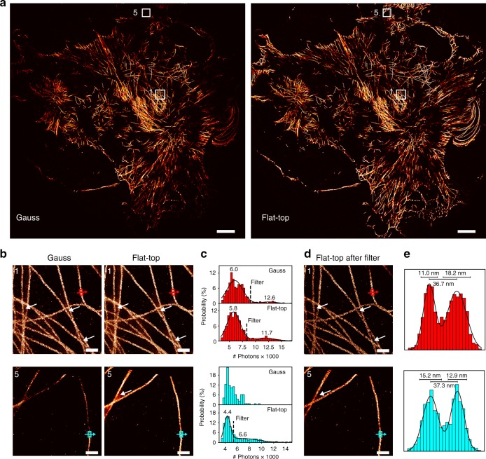
Artifact removal for uniform and quantitative cellular DNA-PAINT imaging. a Full camera chip (130 × 130 µm2) DNA-PAINT image of the microtubule network in fixed COS-7 cells acquired using Gauss illumination (left) and the same field of view for flat-top illumination (right). b Magnified sections from segment 1 and segment 5 (as defined in Fig. 2a) highlighting the image quality in the center and the border region of the camera chip. White arrows point to artifacts due to multi-emitter mislocalizations. c Photon count histograms for box regions in images from b. Double Gaussian fit allows identification and removal of multi-emitter mislocalizations (threshold at 1/e2 of first peak) except for segment 5 for Gaussian illumination. d Filtered flat-top images from b displaying enhanced image quality after removing mislocalization artifacts. e Intensity profiles across single microtubules indicated in d. Scale bars, 10 µm in a, 500 nm in b, d

