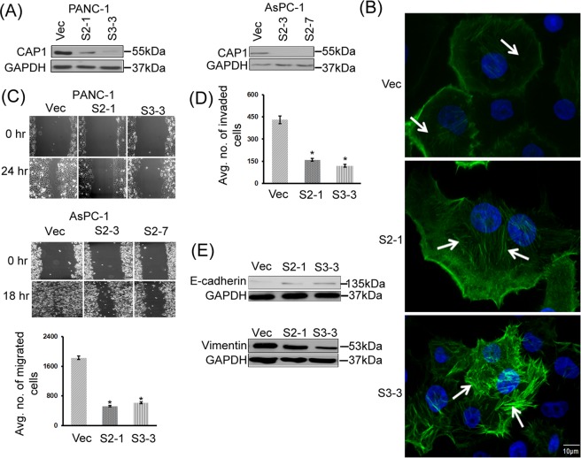
Figure 2.
Knockdown of CAP1 in cancer cells led to enhanced actin stress fibers, and reduced cell motility and invasion. (A) Efficient CAP1 knockdown in two stable clones each for both PANC-1 and AsPC-1 cells as confirmed in Western blotting. CAP1 was detected in Western blotting, where GAPDH was used as a loading control. (B) Depletion of CAP1 in PANC-1 cells led to enhanced actin stress fibers. In control cells that harbor the empty vector for the shRNA (Vec), there were very limited actin stress fibers (indicated with arrows). In contrast, in the CAP1-knockdown cells (S2-1 and S3-3), actin stress fibers were enhanced (indicated with arrows) as observed in fluorescent microscopy following Phalloidin staining. (C) Depletion of CAP1 reduced motility in both PANC-1 and AsPC-1 cells as detected in the wound healing and Transwell migration assays (for PANC-1 only). For Transwell migration assays, ~2 × 10⁴ cells serum-starved overnight were loaded onto each insert placed in a well loaded with medium containing serum. After 16 hrs, migrated cells were scored and data from three independent experiments were collected, analyzed using Student’s t-test, and plotted in the graph where error bars represent S.E.M. “*” indicates P < 0.05 as compared to that in the control cells harboring an empty vector (Vec). (D) Depletion of CAP1 reduced Matrigel invasion in PANC-1 cells. The assays and data collection and analyses were conducted similarly to those in the Transwell migration assays, except that the insert membranes were pre-coated with Matrigel. “*” indicates P < 0.05 as compared with that of the control cells. (E) Reduced EMT in the CAP1 knockdown PANC-1 cells, as indicated by increased E-Cadherin expression as well as reduced Vimentin expression levels in the CAP1-knockdown stable clones. GAPDH serves as a loading control in Western blots.

