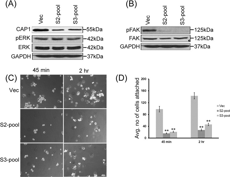
Figure 7.
CAP1-knockdown PANC-1 pool cells had reduced FAK activity and cell adhesion, but no alterations in ERK. (A) CAP1-knockdown PANC-1 pool cells, derived from both S2 and S3 shRNA constructs, did not show altered ERK expression or activity compared with the control pool cells harboring an empty vector, as detected in Western blotting. ERK activity was detected in Western blotting using a phosphor-specific antibody against the Thr202/Tyr204 sites. GAPDH served as a loading control. (B) Reduced FAK activity was detected in the CAP1-knockdown PANC-1 pool cells as compared with the control pool cells. FAK activity was assessed by phosphorylation at Tyr397, using a phosphor-specific antibody against the site in Western blotting; (C) CAP1-knockdown pool cells had reduced cell adhesion at the tested time points (45 minutes and 2 hrs) following plating cells onto fibronectin-coated plates. Approximately 2 × 10⁴ cells were plated onto each well of a 6-well plate, and cells not attached at the indicated time points were washed off. The number of attached cells was counted in five random fields under a phase microscope and images taken. (D) Data collected from three independent cell adhesion assays were analyzed using Student’s t-test, and plotted on the graph where the error bars represent standard deviation. “**” Indicates P < 0.01 as compared to the control cells harboring the empty vector.

