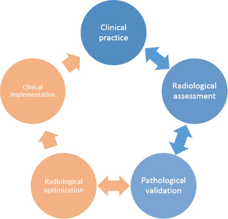
Fig. 1.
Cycle for integration of radiological and pathological development into clinical research and practice. Clinical practice uses radiological assessment (MRI) to assess structural brain changes in patients with neuroinflammatory and neurodegenerative disorders. Initial post-mortem MRI and histopathological studies focus on validating MRI sequences for their pathological sensitivity and specificity (blue). However, more recent studies have focused on optimizing and developing MRI sequences to define more sensitive measures to improve diagnostic and prognostic diagnosis in the clinical setting (orange). As such, post-mortem MRI is of high interest to translate MRI features into histopathological terms but also to translate knowledge on the type or distribution of pathological lesions to MRI, thereby integrating clinical practice, radiological development, and pathological validation to establish optimal radiological biomarkers.

