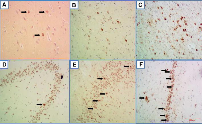
Fig. 5.
Phosphorylated TDP-43-positive structures in the limbic system of AD and PART cases at different Braak stages. A A few NCIs (arrows) in the amygdala of an pre-AD case at stage III. B Some neurofibrillary tangle-like structures in the amygdala of an AD case at stage IV. C Massive NCIs and dystrophic neurites (DNs) in the hippocampus of an AD case at stage VI. D One NCI in a granule cell of the dentate gyrus of a PART case at stage IV. E Some NCIs in the dentate granule cells of an AD case at stage IV. F Massive NCIs in granule cells of the dentate gyrus of an AD case at stage VI. Scale bar, 200 μm.

